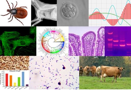
Data collection and analysis in basic, veterinary, and animal science most often requires the use of digital imaging. Moreover, scientists communicate their work to their peers and to non-scientist by means of images. Therefore, researchers must know all the necessary tools to properly acquire and process their images, as well as to build figures that effectively illustrate their scientific work. In addition, in the clinical field, the researchers must be aware of the technical parameters of image acquisition that critically impact the qualitative and quantitative characteristics of anatomical structures and pathology. Such parameters must be properly reported to achieve maximum transparency. During this course the PhD students will learn all the necessary steps that lead to the creation of a figure to be published in a scientific journal in their specific field of interest. The course will cover ground from the acquisition to the submission of the figure, conforming them to publisher’s requirements while ensuring image integrity and transparency.
Practical sessions include acquisition of digital images with different microscopes or devices in basic research as well as in a clinical imaging. Students will decide which session to attend according to their research focus.
Structure of the course
1) Basic principles of Image integrity in scientific publication:
- Overview of the editorial process: how to get your data published in a peer reviewed scientific journal.
- The point of view of the Researcher.
- The point of view of the Editor and the Publisher: what an editor would (and would not) like to receive as an image to be published.
- The point of view of the Reviewer.
- Image integrity in scientific research in basic, veterinary and animal science
2) Critical steps in digital imaging acquisition:
- Image acquisition and processing for scientific research in basic, veterinary and animal science
- Main acquisition parameters of different imaging techniques (X-ray, US, CT, NM, MRI) of medical images required by leading clinical imaging journals
3) Hands on training: image acquisition
- Image acquisition with microscopes and digital cameras (*)
- How to handle graphs and diagrams to be used in scientific publications (1h)
- Image acquisition in a clinical setting
* Students will decide which session to attend according to their research focus.
4) Hands on training: Image processing with GIMP and Adobe Photoshop
- Processing a scientific image with GIMP and Adobe Photoshop
- Managing graphs and vectorial images with GIMP and Adobe Photoshop
- How to build a Figure, composed of images and graphs, to be published in a Scientific Journal
Final examination
The subject of the final exam will be the preparation of a figure to be published in a scientific journal according to the editorial guidelines. Certificate of course completion will be issued.
Professor:
Valentina Lodde
Davide Danilo Zani
Donatella De Zani
Class dates and times:
- March 10 from 9:00am-13:00pm – 14:00pm-18.00 Basic principles of Image integrity in scientific publication & Critical steps in digital imaging acquisition
- March 11 from 9:00am-13:00pm – Research Laboratory (RedBioLab) – Hands on training:image acquisition
- March 12 from 9:00am-13:00pm – Hands on training:image acquisition
- March 18-19 and 20 from 9:30am-12:30am – LABINF03 – Hands on training:image processing

